標題をシェアするに際し、BEYOND ACHONDROPLASIAの投稿を引用・翻訳したいと思います。子どもの軟骨無形成症患者にとって、画期的なセラピーだと思います。皆様の療法士さんにこのビデオを是非見てもらって、一緒に取り組んでもらえれば幸いです。(これは、とっても楽しい日々を過ごしたAISACの年次会合(イタリア・リミニ)での思い出の一つです)
I would like to translate, quote and share the post of “BEYOND ACHONDROPLASIA”. I think it was a really nice program for children with ACH. Please have your therapist see this video and try it together. (It was the one of the great moments during the AISAC congress in RIMINI)
===============
BEYOND ACHONDROPLASIA
http://beyondachondroplasia.org/blogue/?p=6956&lang=en
Mauro Banfiは、フランス マルセイユのOsteopathique de Provence大学でオステオパシーの学位を取得した理学療法士です。軟骨無形成の情報収集と研究を行っているイタリアの団体AISACでMauroに会い、MauroはAISACでオステオパシーによる中耳炎の予防方法を紹介し、その後のワークショップで軟骨無形成の子供に対するいくつかのセラピーを紹介しました。ここでは軟骨無形成の中耳炎を防ぐためにオステオパシーを適用したMauroのビデオを紹介しています。
Mauro Banfi is a physiotherapist and also has a degree in osteopathy, from the College Osteopathique de Provence – Marseille, France. I meet him recently during the AISAC congress. AISAC is the Italian Association for the Information and Study of Achondroplasia. In this event, Mauro presented a proposal for the prevention of otitis media by using osteopathy techniques and after, he did a practical workshop, explaining some of the techniques for young child with achondroplasia. Here is a shared video of Mauro Banfi where he applies osteopathy techniques to prevent otitis media in achondroplasia.
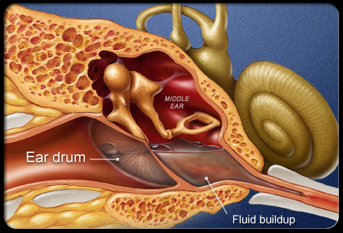
耳管は鼻咽頭と中耳を結ぶチューブ(排水路のようなもの)です。次の図は鼻腔の奥にある耳管が開いている状態を確認する事ができます。分泌物はチューブを通って中耳から食道に排出されます。
子供は耳管が水平的で短く、特に軟骨無形成の子供は短く小さいとされています。軟骨無形の子供は筋肉の低緊張が見受けられる為、1歳くらいまではベッドで横になっていることが多く耳管が一般的な子供に比べ水平的になっていることが多いです。この為、軟骨無形成の中耳炎になりやすく中耳に水が溜まりやすい状態となります。(Benedetto Nicoletti, Elio Ascani, Victor A. McKusick, Human Achondroplasia: A Multidisciplinary Approach, 2012)
The Eustachian tube, also known as the auditory tube or pharyngotympanic tube, is a tube (like a draining channel) that links the nasopharynx to the middle ear. In the next image, you can observe where the Eustachian tube opens (entrance to auditory tube), in an area in the back of the nasal cavity. The secretions that come from the middle ear through the tube are drained to the oesophagus.
In children, the Eustachian tube is horizontal and shorter and “… in a child with achondroplasia, is also short and small. Because these children are quite hypotonic, they may spend the first year of life lying in bed, meaning more pronounced horizontal position that leads to the marked increase of otitis media and the accumulation of fluid in the middle ear seen in childhood in achondroplasia”. (Benedetto Nicoletti, Elio Ascani, Victor A. McKusick, Human Achondroplasia: A Multidisciplinary Approach, 2012)
耳管は骨と軟骨からできており、中耳付近のチューブは骨により構成されています。通常時、チューブは閉じた状態ですが何かを飲み込んだ時や陰圧がかかった際にチューブが開くことになります。
The Eustachian tube consists of a bony part and a cartilaginous part. A portion of the tube (1/3) proximal to (same as next to) the middle ear is made of bone. Usually, the tube is collapsed (closed), but it opens when swallowing and with positive pressure.
耳管には3つの生理的機能があります。
– 耳管の換気と圧力を調整する機能
– 鼻咽頭の分泌物と音の圧力から中耳を保護する機能
– 鼻咽頭へ中耳の分泌物の除去と排出する機能
The eustachian tube has three physiologic functions:
– Ventilation and pressure regulation of the middle ear
– Protection of the middle ear from nasopharyngeal secretions and sound pressures
– Clearance and drainage of middle ear secretions into the nasopharynx
軟骨構造を引っ張る筋肉は、軟骨構造の収縮により定期的に耳管を開口していますが、軟骨無形成の子供のこの筋肉は他の子供のような自然な働きがありません。耳管の自然な開口ができず、耳の感染や中耳に水が溜まることになります。これは耳管を正しく開口できていない為であり、主に炎症、感染、痛み、圧力の上昇、鼓膜の破裂(鼓膜の膨らみ)や熱を引き起こす事になります。
Specific muscles (levator veli palatini, tensor veli palatini) cooperate to open the tube regularly with a traction on a cartilaginous structure (these muscles pull the cartilage structure). But in a child with achondroplasia, these muscles do not have the same efficacy: the Eustachian tube does not accomplish an active natural opening and this leads to ear infections and fluid build-up in the middle ear cavity. This occurs because the tube does not open appropriately, causing mainly inflamation and infection, pain, rise of pression, tympanic membrane rupture (bulging eardrum ) and fever.

Image: Eurolab

This is a tympanic membrane (eardrum) in tension, with fluid inside (the fluid is in the middle ear cavity). Image taken from the ear canal (outside to the inside). Credits: Eurolab.
慢性的な中耳炎の多くは耳鼻科により鼓膜切開を行う事になります。つまり中耳の感染による排液を可能にする為に鼓膜の適切な位置に換気チューブを取り付けます。
In many cases of chronic otitis media, an ENT doctor has to perform a myringotomy, which means to place a ventilation tube, in a precise location of the tympanic membrane (eardrum) to allow drainage of fluid middle ear infection caused by.

Image: ear nose and throat doctor.net
次のビデオでは軟骨無形成の中耳炎の頻度を軽減する別の方法を紹介しています。
Point 1. これは予防であり、中耳炎の兆候がある前にこれを実施する必要があります。次のビデオでは、Mauroが中耳の自然な排液を可能にする為に耳管を開口させるストレッチングセラピーを行っています。
Point 2. 専門家の監督がない状況下では、自分自身でこのセラピーを実施しないようお願いします。最終的にはMauroはこのセラピーの評価についてあなたの理学療法士やオステオパスから問い合わせを受けることも可能です。
Point 3. これらは耳鼻科のアプローチによる補完的なセラピーとなります。
Point 4. 発音する音によってはチューブの開口を促すことが可能です。K, GHやE, Iといった言葉を一緒に遊びながら発音したり、歌ったりしながら、これらの音の発声を子供に教えることができます。これは医療行為ではない為、簡単に実施することができます。
In the next video, we will sho you another way to reduce the frequency of otitis media in achondroplasia.
Point 1. The first idea is that Prevention is the key. So, action must be taken before the signs of otitis occur. In the next video where Mauro Banfi executes streeching techniques in a way to open the eustachian tube, allowing natural drainage of the middle ear.
Point 2. Please, never try to perform these techniques by yourself without professional supervision. Ultimately, you may be refered to Mauro Banfi by your physitherapist or osteopath for evaluation of clinical cases.
Point 3. These techniques are complementary to ENT approach
Point 4. By pronouncing certain sounds, it is possible to stimulate the opening of the tube. You can teach the child to produce words that contain the sounds K and GH, E and I by playing and singing together, which is easy and not invasive.